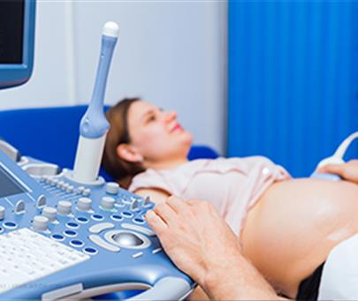
Third-trimester ultrasound is performed at 28-30 weeks of pregnancy and its purpose is to check and find anomalies in the fetus, which could not be identified during the second-trimester screening. 38.5% of anomalies are detected in the third-trimester screening and this group of anomalies is called Late Onset Anomaly, for example, about 40% of urinary system anomalies can be detected after 24 weeks. Also, the anomalies of the digestive system of the fetus can generally be examined and identified after the 24th week of pregnancy (of course, anomalies such as umbilical hernia or gastroschisis can be examined and identified more in the screening of the first trimester). Diseases related to the bone system of the fetus (dysplasia of the bones) can generally be detected after the 24th week of pregnancy (of course, its severe forms cause stillbirth or the birth of a baby with low breathing capacity, which leads to death after birth. can be identified during the screening of the second trimester).
Ultrasound in the third trimester helps check the course and progress of minor anomalies. Mild anomalies of the urinary or digestive system, or even mild heart anomalies and non-fatal bone dysplasia, which have been diagnosed in the previous weeks, should be evaluated by ultrasound and in the form of a specific protocol, and the third time third-trimester ultrasound should be performed in this category. Anomalies are strongly emphasized. The course and progress of such minor anomalies are very important because it gives the sonographer and the parents a good understanding of the course of the anomaly and helps the parents decide on prenatal (during pregnancy) or postnatal (after pregnancy) treatments. is a doer Things such as the evaluation of the placenta in terms of adhesiextractra) are mainly investigated in the screening ultrasound of the third trimester of pregnancy. The normal placenta adheres to the uterine wall and does not invade the myometrium, during delivery the placenta is separated from the decidua by a sudden interruption of intraplacental blood flow at the same time as the contraction of the myometrium.
A placenta that is abnormally attached to the uterine wall after delivery is called placeaccretereta. Placenta accreta is associated with serious complications of pregnancy, including loss of maternal blood, the need to remove the uterus, and remaining remnants of pregnancy products. Placenta accreta can be detected prenatally with ultrasound; So that determining the delivery method can be planned and improve the outcome for mother for baby.
Also, in the screening ultrasound of the third trimester, we get a better understanding of how the fetus grows, and by comparing the findings related to the way the fetus grows (biometrics) with previous ultrasounds, we can make a small estimate about the way the fetus grows and, if necessary, recommend to Color doppler examination of the fetus, placenta and blood supply from mother to placenta and from placenta to fetus. For example, in intrauterine growth retardation (IUGR) with increasing gestational age, diagnosis is achieved by comparing fetal biometrics with fetal growth criteria in previous ultrasounds, and the type of IUGR (relative and non-relative form) is determined by examining the above criteria. And it is very important to perform a color doppler ultrasound in the non-similar form of IUGR, and the doppler criteria indicate the severity of IUGR, how to weigh the fetus, and also the decision to terminate the pregnancy and give birth to the fetus before the due date (term).

